

Case Study 1:
Patient with Neck Pain
Background:
Pancoast tumors, also called superior sulcus tumors, are a rare type of lung cancer that invade the apical chest wall. These tumors can be challenging to identify in practice and are often misdiagnosed, as their symptoms often resemble benign musculoskeletal conditions. Pancoast tumors may invade the brachial plexus, sympathetic chain, and/or vertebral bodies, causing a constellation of symptoms termed Pancoast syndrome, which may include spinal, shoulder, and upper extremity pain and/or weakness, as well as Horner’s syndrome. The most common initial symptom of Pancoast tumors is shoulder pain.
Chiropractors only rarely encounter patients with undiagnosed cancer, yet these patients are important to identify and refer for medical care. In a 2012 survey of chiropractors in the United States, respondents reported encountering a patient with undiagnosed cancer about once every 8 years on average. In another United States survey from 2014, chiropractors reported seeing patients with a bone tumor or metastasis less than once per year.
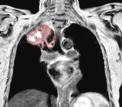

In the Hong Kong Special Administrative Region (HKSAR), the setting of the current case, it is unclear if chiropractors encounter a relatively higher or lower rate of cancer patients than in other countries. However, the incidence of tuberculosis in HKSAR is 10 times greater than that Western developed countries, with 0.6 of every 1000 persons developing tuberculosis per year. Importantly, as illustrated in the current case, tuberculosis is a significant independent risk factor for lung cancer (hazard ratio of 1.6).
Given the potential for chiropractors to rarely encounter patients with undiagnosed cancer, we present a case of Pancoast tumor invading the spine, which was first identified in a chiropractic office.
Patient Details:
Jim, a 59–year-old Asian man presented to a chiropractic clinic in HKSAR with a 1–month history of aching and numbness of his posterior neck and bilateral shoulders. He had an active 6–month history of tuberculosis, which caused an occasional productive cough, and was under medical treatment with a spectrum of antibiotics during this time (ie, isoniazid, rifampin, pyrazinamide, and ethambutol), as supervised by a specialty chest hospital with monthly follow-up visits and serial chest radiographs every 1–2 months.
The patient’s tuberculosis was first suspected 6 months prior after he developed a cough and underwent chest radiography. This was followed up by a Mantoux test, which was strongly positive. His previous providers also initially scheduled a flexible fiberoptic bronchoscopy, but later canceled it as his sputum culture was positive for Mycobacterium tuberculosis, and chest computed tomography (CT) findings were compatible with tuberculosis. At this point he began pharmacologic treatment for tuberculosis with directly observed therapy, which continued through presentation to the chiropractic office.
He described that the neck pain began secondary to episodes of coughing and had negatively impacted his sleep quality. The patient reported an 8 out of 10 pain severity on the numeric rating scale. He had a 50–pack-year history of smoking, was a social drinker, and worked as an interior decorator. The patient had a medical history of hereditary elliptocytosis, and at age 33 had a spontaneous pneumothorax. He denied having a history of cancer. His family history was significant for lung cancer (mother) and hereditary elliptocytosis (father). The patient endorsed occasional hoarseness but denied any bowel or bladder symptoms.
Previous treatment for his neck pain directed by his primary care provider included prescription pain medications (nonsteroidal anti-inflammatory drugs and acetaminophen) and rehabilitation exercises provided by a physical therapist. The patient also had received acupuncture for his neck pain. As no previous treatments provided the patient relief, he sought out chiropractic care (Figure 1).
The patient’s most recent chest radiograph report from 1 month prior reported patchy airspace opacities with a nodular appearance in the right upper lung zone, suggestive of pneumonia. Review of a report of his high-resolution computed tomography (CT) of the thorax from 6 months prior noted right apical fibrocalcific changes, mild bronchiectasis, tiny nodules up to 2 millimeters in both lungs, and shotty non-enlarged mediastinal and hilar lymph nodes. There was no mention of a suspicious lung mass or tumor in these reports.
Your Task:
Task 1:
Send an email to Dr. Joy providing the summary of the patient’s condition, copy her CA, Maria and ask Maria to pull up the CAT Scan of the patient including other possible documentations that will help Dr Joy’s assessment with the patients condition prior the consultation.
Task 2:
Schedule the patient with the CA for an XRay appointment to the nearest Monday from today. Inform the patient that this is part of the routinary check and they need the result prior their second consultation
Task 3:
Call the patient’s insurance provider, Blue Shield and ask if the consultation and XRay exam is coverable.
Task 4:
Once tasks are done, call the patient to book their second consultation reiterating the XRay result should be available.
For questions, reach out to: hanna@chiromatchmakers.com
For systems and tools requests, reach out to: help@chiromatchmakers.com