

Case Study 5:
Elongated Styloid Processes …
Background:
Neck pain is a common problem in the general population with a 12-month prevalence between 30% and 50% and a lifetime prevalence exceeding 60%. [1] Because of its frequency and often-times associated disability, people seek relief of their neck pain from a variety of health care providers. [2] Cervical spine manipulation and mobilization is a common procedure employed by chiropractors, physical therapists, osteopaths, and others to relieve neck pain. [2] Anatomical variations in the neck may present relative contraindications for various types of manipulative procedures, such as high velocity, low amplitude thrust manipulation.
Elongated styloid processes (ESP) and calcified stylohyoid ligaments (CSL) may be part of a condition known as Eagle syndrome. Named after prominent otolaryngologist Dr Watt Eagle [4], Eagle syndrome includes a sensation of a foreign body in the throat, odynophagia, and dysphagia and often includes pain in the neck or face, [3] as cranial nerves are affected by crowding or compression by the anomalous ESP or CSL. Eagle syndrome may also include symptoms suggestive of cerebrovascular compromise, such as dizziness, headache, syncope, and transient visual loss if the internal carotid artery is affected. [4] Symptoms may be more bothersome with head and neck rotation as the structures undergo increased compression or tension from the ESP or CSL. [4]
There is literature that discusses the relevance of ESPs and CSLs to both traumatic and spontaneous internal carotid artery dissection. However, there are few publications that discuss the relevance of these neighboring anatomical structures as they pertain to manual therapies, such as manipulation or mobilization, of the cervical spine. The purpose of this paper is to present a case of a patient with neck pain, tinnitus, and headache in the setting of bilateral ESPs and CSLs and how knowledge of this anatomical variation and symptomatic presentation affected the rehabilitation management plan for this patient. We also discuss the potential relevance of ESPs and CSLs to carotid artery dissection.
Patient Details:
Steve, a 29-year-old male United States Marine helicopter mechanic was referred to the primary author for chiropractic care for chronic pain in the right side of his neck and upper back, which started 7 years earlier after sustaining a cervical flexion injury during Marine Corps martial arts training. At that time he fell from a height of approximately 3 ft onto the back of his head. The patient described the chronic, constant pain as an aching sensation and he felt the need to frequently stretch his neck or “crack it”; however, nothing actually provided sustained relief. Prolonged static postures, such as sustained desk work, or vigorous activity tended to aggravate the pain. He had no pain or altered sensation in his upper extremities. His pain was generally moderate in intensity, rating it a 7 out of 10 on a verbal pain scale (VPS). He had received various interventions over the previous several years from his primary care providers, neurologists, physical therapists, and chiropractors without resolution. A review of systems revealed that the patient had concurrent tinnitus and dizziness and a past history of right side parietal headaches and right side tonsillitis. He had no loss of consciousness, no dysphagia, dysphonia, nystagmus, nausea or vomiting.
The patient posture demonstrated anterior head carriage, increased kyphosis and rounded forward shoulders. He had an increase in neck and upper back pain on his right side with flexion and left rotation of his neck. The patient’s neck pain was reproduced by pressing down on the top of his head and palpation of the right cervicothoracic extensor and upper trapezius muscles. He showed no signs of cervical nerve root tension or impingement. Cranial nerve, cervical nerve root, and brachial plexus examinations were unremarkable.
The rehabilitation regimen was created using precautions to avoid end range loading of the cervical spine into rotation, lateral bending, or combinations of the 2 movements due to the ESP/CSL. The upper thoracic spine was manipulated in the prone position while the head of the patient was stabilized to reduce rotation and lateral bending and the head portion of the table was set to maintain spinal neutral. Supine or seated cervical spine manipulation procedures were not used to avoid rotating the cervical spine. Soft tissue mobilization of the right cervical paravertebral and upper trapezius muscles was performed with the patient in a seated position. For these procedures, the patient positioned his head into the contracted muscle position and then slowly moved to the neutral cervical position while digital pressure was applied to tender points in the muscle. The patient was also given a home exercise program of corrective postural exercises that included stretching, strengthening, and proprioceptive exercises. Education on proper computer ergonomics was also provided since part of his job involved prolonged computer use.
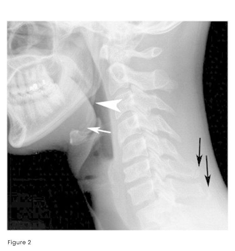
Conventional radiographs had been previously obtained by the patient’s primary care provider 1 month prior to his chiropractic consultation and demonstrated old C6 and C7 spinous process (“clay shoveler’s”) fractures, degenerative disc disease at C6/7 hypolordosis, and a right ESP with left CSL (Figure 2). The radiologist noted that ESPs and CSL can be seen in Eagle syndrome and recommended clinical correlation. Thoracic plain films were normal. The patient was diagnosed with mechanical cervicothoracic pain and myalgia.
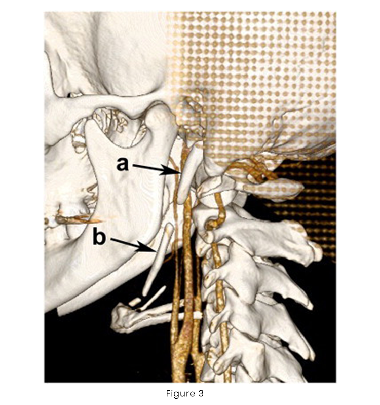
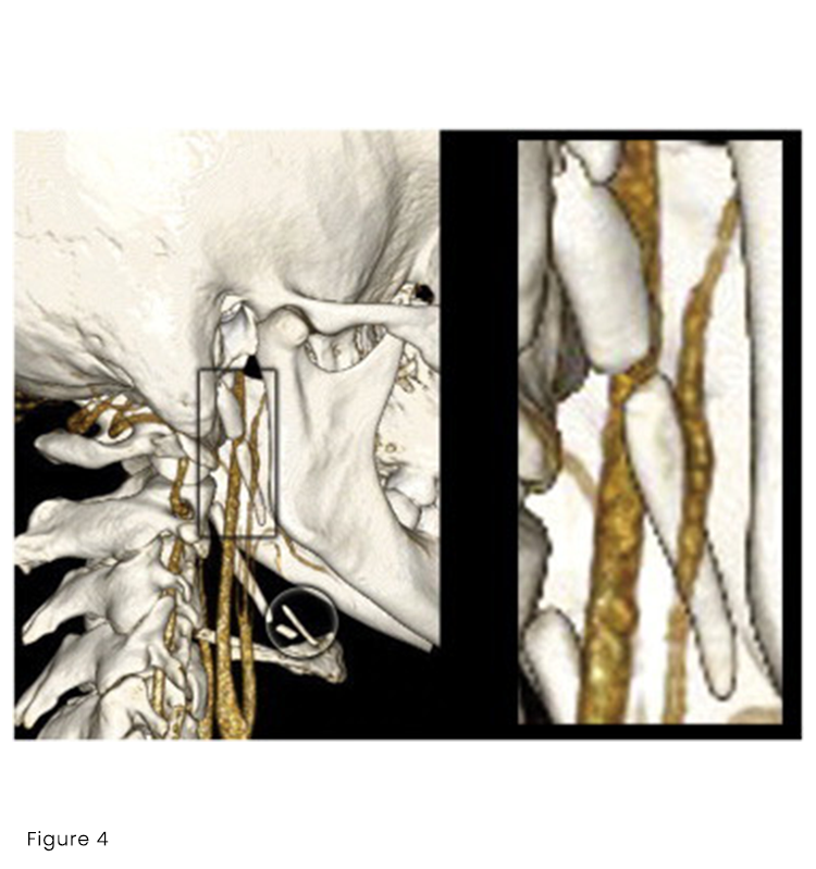
At his third office visit he stated that his tinnitus was more predominant in his right ear but denied any alteration in symptoms with changes in head position and no positional dizziness, which are both characteristic of Eagle syndrome. He had no headache on this visit and his neck pain severity was reduced to a VPS of 4/10) from previous visits. A re-examination revealed no significant changes in his exam findings. With Eagle syndrome being a differential diagnosis, specific confirmatory tests were conducted, which were negative, including no reproduction of Eagle syndrome symptoms with palpation of the tonsillar pillars, and no increase in tinnitus or production of dizziness with active or passive head positioning into end range of motion for extension, flexion, side bending, or rotation. Treatment was continued and computed tomography angiography of the neck was ordered after consultation with the radiologist. The patient’s pain plateaued by the fifth office visit. He reported mild reduction in his pain after each treatment but the pain would return to moderate levels of severity within 24 to 48 hours. The patient’s tinnitus also plateaued. Treatment focusing on a home based exercise program for postural correction continued while awaiting the computed tomography examination and results. The volumetric helical computed tomography demonstrated calcification of the stylohyoid ligaments and imaging of the spatial relationship between the ESPs and carotid arteries (Figures 3-4). At the report of imaging findings the patient reported no improvements in his symptoms and an elevated level of concern regarding his tinnitus. A consultation with an otolaryngologist was made for a definitive evaluation for Eagle syndrome or other pathology and the patient was also referred to a physical therapist for acupuncture treatment while he continued with chiropractic care.
The patient was thoroughly evaluated by the otolaryngologist to include a comprehensive series of hearing tests. His physical examination was normal and he had normal tympanograms. His hearing tests showed mild high frequency bilateral hearing reduction that was symmetrical and consistent with occupational exposure to loud noises. The otolaryngologist reported that the patient did not have Eagle syndrome.
The patient had a total of 8 chiropractic treatments and 5 physical therapy (acupuncture) treatments. He reported decreased neck stiffness but his pain remained at a level of approximately VPS of 4/10. He continued to have tinnitus. No adverse effects were reported during treatment. Due to military commitments and a lack of further improvement in his symptoms, care was discontinued.
Your Task:
Task 1:
Send an email to Dr. Joy providing the summary of the patient’s condition, copy her CA, Maria and ask Maria to pull up the Scan of the patient including other possible documentations that will help Dr Joy’s assessment with the patients condition prior the consultation.
Task 2:
Schedule the patient for a twice a week for 2 weeks to include 4 acupuncture treatments with needles placed as indicated by clinical exam but usually in the above locations. acupuncture will include treatment consisted of electroacupuncture for 20 minutes at 2 Hz with needles placed at the right BL11, TH15, BL42, and BL44 as well as GB20 and GV20 without electrical stimulation.
Task 3:
Schedule patient with additional needle placement without electrical stimulation of the right Shen Men point and point zero. The patient’s numerical pain scale usually was a 4/10 before acupuncture treatment and either the same or 1 point lower after treatment.
Task 4:
Once tasks are done, send an email to the patient and discuss the recurrence of their chiropractic sessions starting 2nd week of next month.
For questions, reach out to: hanna@chiromatchmakers.com
For systems and tools requests, reach out to: help@chiromatchmakers.com